
In cellular bioenergetics, the traditional view of mitochondria has centered almost exclusively on their role as ATP-generating organelle powerhouses. However, modern molecular biology has revealed that these organelles also function as active signaling hubs. Mitochondria communicate dynamically with the nuclear genome through retrograde signaling mechanisms, releasing bioactive peptides encoded directly within their own circular mitochondrial DNA (mtDNA).
Among these mitochondrial-derived peptides (MDPs), MOTS-c (Mitochondrial Open Reading Frame of the 12S rRNA Type-c) has emerged as a critical regulator of metabolic homeostasis, insulin sensitivity, and cellular stress responses. Synthesized as a 16-amino-acid sequence ($Met-Arg-Trp-Gln-Glu-Met-Gly-Tyr-Ile-Phe-Tyr-Pro-Arg-Lys-Leu-Arg$), MOTS-c translocates to the nucleus under metabolic stress, binding directly to DNA response elements to alter nuclear gene expression.
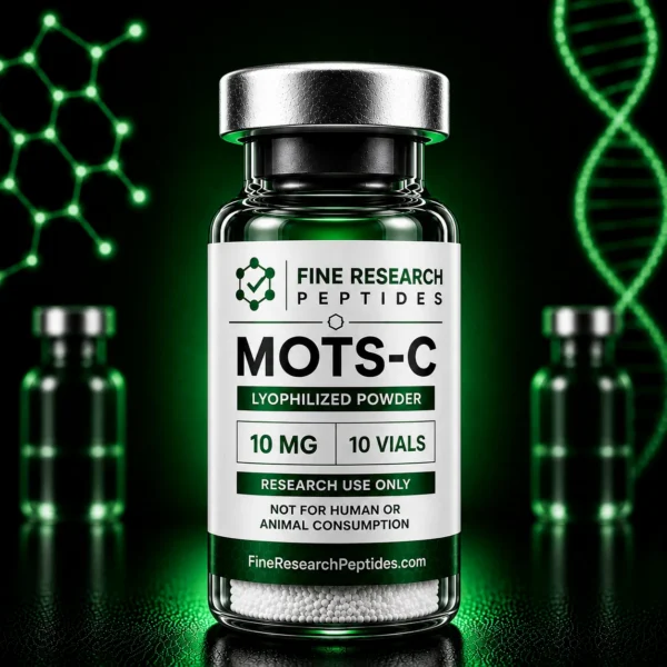
For research facilities, academic laboratories, and contract research organizations (CROs) designing metabolic studies, establishing standardized experimental protocols around a high-purity mots-c 10mg vial configuration is essential. Utilizing pristine starting materials ensures that longitudinal bioenergetic assays yield clean, reproducible, and publication-grade datasets.
1. The Retrograde Signaling Mechanism: From Mitochondrion to Nucleus
Understanding the unique biochemical role of MOTS-c requires analyzing its retrograde signaling path. While most mitochondrial proteins are encoded by nuclear DNA and imported into the organelle, MOTS-c is encoded within the 12S ribosomal RNA region of the mitochondrial genome.
The intracellular pathway of MOTS-c involves several key stages:
-
AMPK Activation: Upon exposure to metabolic stressors such as glucose deprivation or exercise-induced energy demands, MOTS-c activates $AMP$-activated protein kinase ($AMPK$). This activation occurs independently of intracellular $AMP/ATP$ ratios through direct inhibition of the folate cycle and $de novo$ purine biosynthesis.
-
Nuclear Translocation: Following $AMPK$ activation, MOTS-c translocates from the cytoplasm into the cell nucleus, a process mediated by stress-sensitive transcription factors such as $Nrf2$.
-
Nuclear Gene Regulation: Inside the nucleus, MOTS-c binds directly to promoter regions containing Antioxidant Response Elements ($AREs$). This interaction upregulates genes involved in lipid oxidation, glucose uptake, and endogenous antioxidant defenses, effectively reprogramming cellular metabolism.
2. Metabolic Homeostasis: Skeletal Muscle and Adipose Remodeling
In preclinical models evaluating metabolic syndrome, type 2 diabetes, and age-related insulin resistance, MOTS-c exhibits profound effects on peripheral glucose clearance and lipid handling.
Key physiological response patterns documented in preclinical MOTS-c models include:
-
Skeletal Muscle Glucose Uptake: MOTS-c stimulates $GLUT4$ glucose transporter translocation to the cell membrane in skeletal muscle myocytes, enhancing insulin-independent glucose uptake even under high-fat diet conditions.
-
Adipose Tissue Browning: The peptide promotes the transdifferentiation of white adipose tissue ($WAT$) into thermogenic, beige-like fat by upregulating uncoupling protein-1 ($UCP1$) expression and mitochondrial density.
-
Systemic Energy Expenditure: By shifting substrate utilization toward fatty acid oxidation and preventing diet-induced weight gain, MOTS-c maintains systemic energy balance without relying on central nervous system appetite suppression.
3. Analytical Purity Standards: Overcoming 16-Residue Synthetic Impurities
MOTS-c is a 16-amino-acid peptide with a hydrophobic core ($Ile-Phe-Tyr-Pro-Arg-Lys-Leu-Arg$). Assembling this sequence via Solid-Phase Peptide Synthesis ($SPPS$) presents specific chemical challenges, including peptide aggregation on the resin matrix and incomplete coupling of hydrophobic residues.
| Quality Parameter | Standard Import Grade | Certified USA Research Standard | Preclinical Impact |
| RP-HPLC Purity Analysis | Variable (85–92%) | Guaranteed $ge$98% per batch | Prevents truncated fragments from obscuring $AMPK$ kinetic curves |
| ESI-MS Mass Verification | Unverified or missing | Confirmed mass ($2174.6text{ g/mol}$) | Validates exact 16-amino-acid sequence assembly |
| Residual Salt Profile | High TFA salt levels | Automated counter-ion exchange | Prevents cell culture toxicity and media pH instability |
| Reconstitution Vector | Unbuffered sterile water | Pure bacteriostatic water for peptides | Halts microbial growth and preserves multi-dose stock solutions |
When research groups prepare to buy peptides online for research use, relying on unverified suppliers risks introducing deletion sequences or unreacted reagents into sensitive cellular assays. Securing materials from a certified research peptide supplier usa ensures that every batch includes lot-matched Reverse-Phase High-Performance Liquid Chromatography ($RP-HPLC$) and Electrospray Ionization Mass Spectrometry ($ESI-MS$) documentation.
4. Reconstitution Safety and Liquid-Phase Preservation
Maintaining the structural integrity of a mots-c 10mg vial following liquid transition is critical for multi-day testing protocols. Lyophilized (freeze-dried) peptide cakes are stable under long-term freezer storage, but once dissolved into liquid media, their peptide bonds become susceptible to hydrolytic cleavage and microbial contamination.
In multi-dose research schedules, accessing a single vial repeatedly introduces airborne bacterial and fungal spores. To protect the sequence against enzymatic decay, laboratory standard operating procedures mandate reconstituting freeze-dried material with high-grade bacteriostatic water for peptides.
Formulated with 0.9% USP-grade benzyl alcohol, fresh bacteriostatic water 10ml or 10ml bacteriostatic water lines prevent microbial growth for up to 28 days under refrigeration ($2^circtext{C}$ to $8^circtext{C}$). This preservation step ensures that stock solutions retain volumetric dosing accuracy and molecular stability across extended research timelines.
5. Endotoxin Management and Environmental Logistics
A major risk in mitochondrial bioenergetic studies is the presence of bacterial endotoxins—lipopolysaccharide ($LPS$) fragments from Gram-negative bacterial cell walls that easily survive basic sterile micro-filtration.
Cellular respiration assays, such as Seahorse XF flux analysis, are sensitive to endotoxin contamination. When introduced into cell culture models, trace endotoxins trigger Toll-like receptor 4 ($TLR4$) signaling, driving inflammatory cytokine release, altering mitochondrial membrane potential ($DeltaPsi_m$), and uncoupling oxidative phosphorylation. These artifacts skew oxygen consumption rate ($OCR$) measurements, rendering experimental data unrepeatable.
Partnering with an authenticated domestic supplier guarantees that research lots undergo Limulus Amebocyte Lysate ($LAL$) testing to confirm endotoxin levels remain safely below 0.25 EU/mg. Furthermore, temperature-monitored cold-chain shipping networks protect heat-sensitive peptides from thermal degradation during transport, delivering intact, fully functional reagents directly to the laboratory bench.
Conclusion: Advancing Mitochondrial Research Through Quality Reagents
As preclinical research continues to illuminate the complex retrograde signaling networks controlled by mitochondrial-derived peptides, high-purity testing reagents remain essential to scientific discovery. Utilizing a fully authenticated mots-c 10mg starting material enables research facilities to evaluate $AMPK$ activation, nuclear gene translocation, and cellular bioenergetics with high precision and reproducibility.
Sourcing inventory through an established research peptide supplier usa eliminates sequence variations, endotoxin contamination, and thermal degradation risks. Paired with proper reconstitution using bacteriostatic water for reconstituting peptides, research teams can build robust experimental frameworks, generate publication-grade datasets, and advance the scientific understanding of mitochondrial metabolic regulation.