Ultrasound technology has revolutionised modern medicine, offering a safe and painless way to examine the body’s internal structures. Using high-frequency sound waves, this imaging technique creates detailed pictures of organs, tissues, and blood vessels. Unlike X-rays or CT scans, ultrasound doesn’t use ionising radiation, making it a preferred choice for many diagnostic situations especially during pregnancy.
In this guide, we’ll explore the various types of ultrasound scans available, from abdominal and pelvic examinations to specialised cardiac imaging. You’ll learn what conditions ultrasound can detect, how to prepare for your scan, and what happens during the procedure. Whether you’re expecting a baby, investigating abdominal pain, or monitoring a health condition, understanding ultrasound technology can help you feel more confident about your upcoming appointment.
Common Types of Ultrasound Scans Explained
Abdominal Ultrasound
An abdominal ultrasound examines organs within your belly, including the liver, gallbladder, spleen, pancreas, and kidneys. Doctors commonly request this scan to investigate unexplained abdominal pain, check for gallstones, or monitor liver conditions.
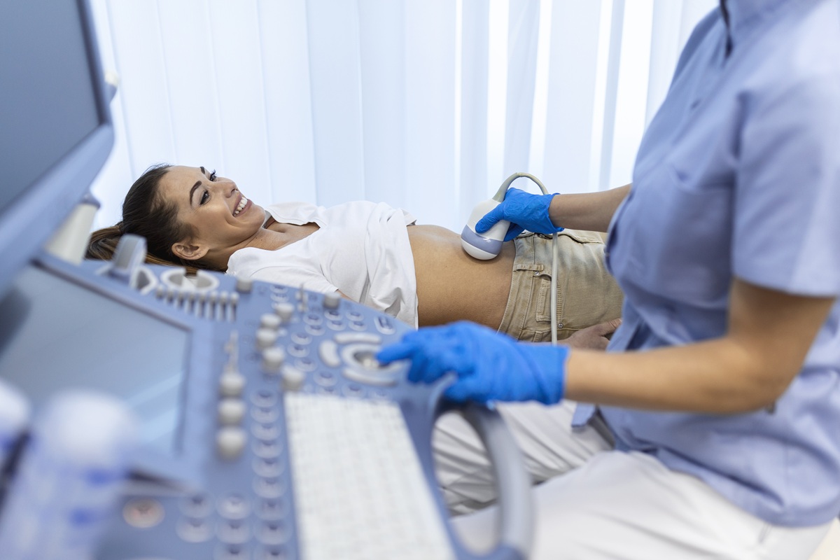
During the procedure, a sonographer applies warm gel to your abdomen and moves a handheld device called a transducer across your skin. The transducer sends sound waves into your body, which bounce back to create images on a screen. You’ll typically need to fast for several hours beforehand to improve image quality.
Pelvic Ultrasound
Pelvic ultrasounds focus on the reproductive organs and bladder. For women, this includes examining the uterus, ovaries, and fallopian tubes. Men may have pelvic ultrasounds to assess the prostate and seminal vesicles.
This type of scan helps diagnose conditions like ovarian cysts, fibroids, and endometriosis. Doctors also use pelvic ultrasounds to investigate abnormal bleeding, pelvic pain, or fertility concerns. You’ll usually need a full bladder for this examination, as it helps create clearer images.
.jpg)
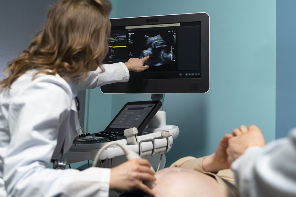
Pregnancy Ultrasound (Obstetric Ultrasound)
Perhaps the most well-known type, pregnancy ultrasounds monitor your baby’s development throughout gestation. Most Australian women have at least two routine scans one around 12 weeks and another between 18-20 weeks.
These scans confirm your due date, check for multiple pregnancies, and assess your baby’s growth and anatomy. They can also detect certain chromosomal abnormalities and structural problems. Many parents treasure these scans as their first glimpse of their little one.
Musculoskeletal Ultrasound
Musculoskeletal ultrasound examines muscles, tendons, ligaments, and joints. It’s particularly useful for diagnosing sports injuries, tendonitis, and soft tissue problems that don’t show up well on X-rays.
This type of scan offers the advantage of real-time imaging, allowing doctors to see how structures move during certain activities. It’s commonly used to guide injections into joints or soft tissues, improving accuracy and effectiveness.
Vascular Ultrasound (Doppler Ultrasound)
Doppler ultrasound measures blood flow through your arteries and veins. It uses a special technique that detects the movement of blood cells, helping identify blockages, blood clots, or narrowed vessels.
Doctors often recommend vascular ultrasounds for patients with circulation problems, leg swelling, or risk factors for deep vein thrombosis. The carotid arteries in your neck are frequently examined using this technique to assess stroke risk.
Cardiac Ultrasound (Echocardiogram)
An echocardiogram provides detailed images of your heart’s structure and function. It shows the heart chambers, valves, and surrounding blood vessels, revealing how well your heart pumps blood.
This scan is essential for diagnosing heart conditions like valve problems, heart failure, and congenital defects. It’s completely painless and typically takes 30-60 minutes. Cardiologists rely heavily on echocardiograms for both diagnosis and ongoing monitoring.
Breast Ultrasound
Breast ultrasounds often complement mammograms, particularly for women with dense breast tissue. They help distinguish between solid masses and fluid-filled cysts, which appear quite differently on ultrasound.
If you’ve noticed a breast lump or your mammogram shows something requiring further investigation, your doctor may request this scan. It’s also useful for guiding needle biopsies when tissue samples are needed.
Thyroid Ultrasound
Thyroid ultrasounds examine the butterfly-shaped gland in your neck. They can detect nodules, cysts, and other abnormalities that might affect hormone production.
Your doctor might recommend this scan if blood tests suggest thyroid dysfunction or if they feel a lump during a physical examination. Thyroid ultrasounds help determine whether nodules require biopsy or monitoring.
What Are the Different Types of Ultrasound Techniques?
2D Ultrasound
Traditional 2D ultrasound creates flat, black-and-white images that trained professionals interpret. This remains the most common technique and provides excellent diagnostic information for most purposes.
3D and 4D Ultrasound
3D ultrasound compiles multiple 2D images to create three-dimensional pictures. 4D adds the element of real-time movement—essentially a 3D video. These techniques are particularly popular during pregnancy for detailed views of your baby’s face and features.
Transvaginal Ultrasound
For clearer views of pelvic organs, a slim probe may be inserted into the vagina. This technique provides more detailed images of the uterus and ovaries than external scanning, making it valuable for early pregnancy assessment and investigating gynaecological conditions.
Transrectal Ultrasound
Similarly, transrectal ultrasound uses a probe inserted into the rectum to examine the prostate gland closely. It’s commonly performed when investigating prostate abnormalities or guiding biopsies.
What Conditions Can an Ultrasound Detect?
Ultrasound can identify a remarkable range of conditions across different body systems. In the abdomen, it detects gallstones, kidney stones, liver disease, and abdominal aortic aneurysms. Pelvic scans reveal ovarian cysts, fibroids, and ectopic pregnancies.
Cardiac ultrasounds diagnose valve problems, heart muscle weakness, and fluid around the heart. Vascular studies identify blood clots, arterial blockages, and varicose veins. Musculoskeletal ultrasounds pinpoint tendon tears, muscle injuries, and joint inflammation.
While ultrasound excels at examining soft tissues, it has limitations. It cannot see through bone or air-filled structures very well, which is why other imaging methods might be needed for certain conditions.
Is Ultrasound Safe? Understanding the Risks and Benefits
Ultrasound is considered extremely safe with no known harmful effects when performed by qualified professionals. Unlike X-rays and CT scans, it doesn’t use ionising radiation, making it ideal for pregnant women and children.
The main benefits include real-time imaging, no recovery time, and relatively low cost compared to other imaging methods. There’s no need for anaesthesia, and most scans cause no discomfort whatsoever.
The only consideration is that ultrasound has diagnostic limitations it may not provide sufficient detail for certain conditions, requiring additional imaging.
How Should You Prepare for an Ultrasound Scan?
Preparation varies depending on which type of ultrasound you’re having. For abdominal scans, you’ll typically fast for 6-8 hours beforehand. Pelvic ultrasounds usually require a full bladder, so drink plenty of water before your appointment.
Wear comfortable, loose fitting clothing that allows easy access to the area being scanned. Avoid applying lotions or powders to the scan area. Bring your referral, Medicare card, and any previous imaging results.
What Happens During an Ultrasound Procedure?
You’ll lie on an examination table while the sonographer applies warm gel to your skin. This gel helps transmit the sound waves between the transducer and your body.
The sonographer moves the transducer across your skin, capturing images and measurements. You might be asked to hold your breath briefly or change positions. The images appear on a screen in real-time, though the sonographer may not be able to discuss findings during the scan.
How Long Does an Ultrasound Take?
Most ultrasounds take between 15-45 minutes, depending on the type and complexity. Simple scans may be completed quickly, while detailed examinations like echocardiograms take longer. You’ll typically be able to resume normal activities immediately afterwards.
When Would a Doctor Recommend an Ultrasound Over Other Imaging?
Doctors choose ultrasound when they need real-time imaging of soft tissues without radiation exposure. It’s the first choice for pregnancy monitoring, examining superficial structures, and assessing blood flow. Ultrasound is also more accessible and affordable than MRI or CT scanning in many situations.
Understanding Your Ultrasound Results: What to Expect
A radiologist analyses your ultrasound images and sends a report to your referring doctor, usually within a few days. Your doctor will then discuss the findings with you and explain any recommended follow-up.
Normal results mean nothing abnormal was detected. Abnormal results require further discussion about next steps, which might include additional testing, monitoring, or treatment.
Frequently Asked Questions About Ultrasound Scans
Can Ultrasound Detect Cancer?
Ultrasound can identify suspicious masses and abnormalities that might indicate cancer. However, a definitive cancer diagnosis typically requires biopsy and laboratory analysis. Ultrasound often guides these biopsy procedures.
What Is the Difference Between an Ultrasound and a Sonogram?
These terms are often used interchangeably. Technically, ultrasound refers to the imaging technique using sound waves, while a sonogram is the actual image produced. In practice, most people understand both terms to mean the same thing.
Choosing the Right Ultrasound for Your Needs
Ultrasound technology offers an incredible window into the human body, providing valuable diagnostic information across numerous medical specialties. From monitoring precious new life during pregnancy to investigating complex cardiac conditions, these scans play an essential role in modern healthcare.
Understanding the different types of ultrasound scans helps you feel prepared and informed when your doctor recommends this imaging. Whether you need an abdominal scan for digestive concerns, a musculoskeletal assessment for joint pain, or routine pregnancy monitoring, ultrasound provides safe, painless, and effective answers.
If you’ve been referred for an ultrasound, don’t hesitate to ask your doctor or the imaging clinic any questions about preparation or what to expect. Knowledge truly is power when it comes to your health, and being an informed patient leads to better outcomes. Your ultrasound is simply a tool to help your healthcare team provide you with the best possible care.