The Breast Imaging Conversation Most Women Never Get to Have
If you’ve ever left a mammogram appointment with more anxiety than answers, you’re not alone. A callback. An unclear result. A recommendation for “additional imaging.” And suddenly what was supposed to be a routine screening feels like the beginning of something terrifying — even when nothing is actually wrong.
Here’s what most women don’t know: the technology available for breast imaging has changed significantly in the last decade, and most of us are still operating on outdated assumptions about what screening looks like, what it feels like, and what it can actually find.
A breast ct scan is one of the most significant advances in breast imaging in recent years, and the majority of women who could benefit from it have never heard of it. That’s worth fixing.
What a Breast CT Scan Actually Is
Let’s clear up the confusion right away, because “CT scan” in the context of breast imaging doesn’t mean the same thing as the full-body CT scans you might have had for other medical reasons.
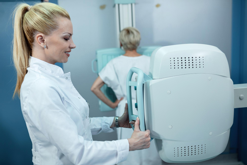
A dedicated breast CT scan — like the Koning Vera system available through Gnosis For Her — is specifically engineered for breast tissue. You lie face down on a padded table, your breast rests naturally in an opening, and the scanner rotates around it in about ten seconds per side. No compression. No squeezing. No breath-holding in discomfort.
The result is a full three-dimensional image of the breast from every angle — not a flattened, compressed 2D view, but an actual volumetric picture that lets radiologists see through and around overlapping tissue.
That matters enormously, especially for women with dense breast tissue.
Why Dense Breast Tissue Changes Everything
Almost half of all women in the United States have dense breast tissue. Dense tissue appears white on a mammogram. So does cancer. On a standard mammogram, dense tissue can quite literally hide what’s there — a phenomenon sometimes called the “masking effect.”
This isn’t a flaw with mammography exactly. It’s a physics problem. You’re compressing a three-dimensional structure into a two-dimensional image, and when the background tissue is dense, the contrast between normal and abnormal disappears.
A breast ct scan solves this by removing the compression and the flattening entirely. The 3D imaging captures each layer of tissue independently, which means dense tissue no longer creates the same visual interference. What was hidden in a conventional mammogram becomes visible in a way it simply wasn’t before.
For women who have been told they have dense breast tissue — often just in a letter after their annual mammogram, with little explanation of what it actually means for their risk — this is a real, meaningful step forward.
Who Should Be Thinking About This Technology
You don’t have to have a prior cancer diagnosis to benefit from a breast CT scan. In fact, many of the women who get the most value from this technology are those in specific situations that conventional mammography handles poorly.
Women recalled after a mammogram
If your mammogram flagged something and you’ve been asked to come back for additional imaging, a breast CT scan is often the clearest next step. Rather than repeat compression views or an ultrasound that still leaves uncertainty, the 3D imaging can provide the kind of detail that leads to an actual answer — and far fewer unnecessary biopsies.
Women with a personal or family history of breast cancer
When risk is elevated, the margin for missing something narrows. Clearer imaging isn’t just reassuring — it’s strategic. For high-risk women, a breast CT scan can be part of a more comprehensive surveillance approach alongside or instead of conventional screening.
Women who find mammography physically intolerable
This one gets talked about less than it should. There is a real population of women — those with hypersensitivity, fibromyalgia, prior surgery, implants, or simply a lower pain threshold — for whom the compression involved in a standard mammogram is genuinely difficult or impossible to endure. No compression breast imaging isn’t just a comfort upgrade. For these women, it’s what makes imaging possible at all.
Women who’ve had inconclusive results
“We need to monitor this” is not an answer. It’s a deferral. If you’ve been living with unresolved uncertainty from a prior imaging result, more detailed 3D imaging can often provide the clarity that ends the waiting game.
The Ten-Second Difference
One of the things that surprises women most when they learn about the Koning Vera system is the speed. Ten seconds per breast. That’s the scan time. Not the appointment — the actual imaging acquisition.
Compare that to the experience most women are used to: repositioning, repeat compressions, holding your breath, the technologist leaving the room, and coming back to adjust the compression plate again. The entire encounter feels drawn out and uncomfortable, and for many women, it’s a reason they delay or skip screening altogether.
When the scan takes ten seconds and doesn’t involve compression, the barrier to showing up is dramatically lower. And in breast cancer detection, showing up is everything.
What “3D” Really Means for Your Results
The distinction between a 2D mammogram and 3d no compression breast imaging isn’t just a technical footnote. It changes what radiologists can see and what conclusions they can draw with confidence.
In a standard mammogram, overlapping tissue structures can create shadows that look like abnormalities — leading to callbacks and additional testing that turn out to be nothing. They can also obscure real abnormalities behind dense tissue — leading to missed findings. Both of these failure modes have real consequences: one causes unnecessary anxiety and procedures, the other causes delayed diagnosis.
3D imaging addresses both simultaneously. By capturing the breast in volumetric slices, it allows radiologists to scroll through tissue layer by layer, separating structures that would overlap in a flat image. The result is fewer false positives — which means fewer unnecessary callbacks — and better sensitivity for real findings hiding within dense tissue.
This isn’t incremental improvement. It’s a fundamentally different tool.
Health Equity and the Mobile Clinic Model
Something that distinguishes Gnosis For Her from a standard imaging center is how the service reaches women. Access to advanced breast imaging has historically been concentrated in affluent zip codes and major medical centers. Women in underserved communities — those without easy transportation, flexible work schedules, or established relationships with specialists — have largely been excluded from the benefits of newer imaging technology.
Gnosis For Her’s mobile clinic model brings the breast CT scan directly into communities across Southern California. The scan itself, including a physician read, is accessible at a price point designed to remove cost as a barrier. For women who’ve never had the conversation about what comes after an unclear mammogram, this is the conversation — and the technology — coming to them.
Stop Waiting for Clarity That Isn’t Coming From the Same Technology
If you’ve had a mammogram result that left you uncertain, if you’ve been told you have dense breast tissue without a clear follow-up path, or if you’ve avoided screening because compression is painful or impossible for you — there is a better option available right now.
A breast ct scan at Gnosis For Her takes ten seconds per breast, requires no compression, and produces the kind of 3D detail that changes what’s visible and what’s knowable about your breast health.
You deserve imaging that actually works for your body. Book your scan today at gnosisforher.com — and get the clarity you’ve been waiting for.